Microscope Cameras
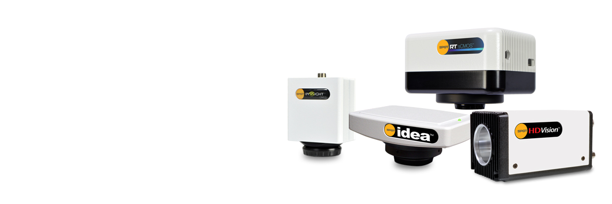
SPOT Cameras
Scientific Digital Cameras for Microscopy
SPOT Imaging manufactures a complete line of scientific digital cameras for microscopy, from easy to use color CMOS cameras for brightfield microscopy to ultra-sensitive scientific CMOS sensors for low light fluorescence applications.
Every SPOT camera comes with the SPOT Software™, an easy to use image capture application full of tools for microscopists, including scale bar, annotations, measurements, custom reporting, and time-lapse recordings. Cameras run on both Windows® and Macintosh® computers, and a Software Development Kit is available for integration with custom software. All SPOT cameras ship with a 2-year manufacturer’s warranty and our exceptional track record for building reliable cameras that last for years.
SPOT Cameras
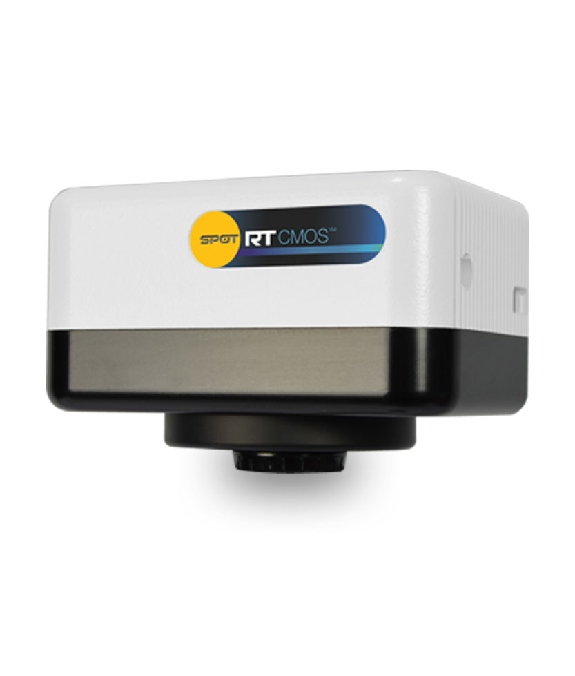
SPOT RT™Cooled sCMOS Camera
The SPOT RT sCMOS uses Sony’s breakthrough Pregius™ CMOS sensor. Now you can experience unprecedented speed and sensitivity in a scientific CMOS camera. Deep cooling allows dim images to be seen without becoming obscured by dark current.
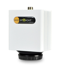
SPOT Insight™ CMOS Cameras
The SPOT Insight CMOS Camera featuring Sony’s Pregius™ CMOS sensor, delivers high speed, high resolution and high QE — features previously associated with much higher-priced cameras. Now available in 5MP and 12MP models!
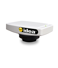
SPOT Idea™ CMOS Camera
The SPOT Idea CMOS cameras deliver high impact color images for journal publication and industrial documentation at an economical price.
SPOT HDVision™ High Definition Video Camera
The SPOT HDVision cameras provide live video microscopy imaging in HD for conferences and classrooms without requiring a computer.
|
Key:* Best Choice for Application √ Capable + Capability Surpasses Application x Not Recommended
|
||||||
| RT sCMOS | Idea | Insight | ||||
|---|---|---|---|---|---|---|
Brightfield Macrophotography Applications |
||||||
| Failure Analysis |
+
|
*
|
*
|
|||
| Inspection |
+
|
*
|
*
|
|||
| Metrology |
+
|
*
|
*
|
|||
| Semiconductor |
+
|
*
|
*
|
|||
| Machine Vision |
+
|
*
|
*
|
|||
| Forensic |
+
|
*
|
*
|
|||
| Brightfield Gels |
+
|
√
|
*
|
|||
Low Light Macrophotography Applications |
||||||
| Semiconductor Emission Analysis |
*
|
x
|
√
|
|||
| Fluorescence Gels |
*
|
x
|
√
|
|||
| Multiplex Gel Imaging |
*
|
x
|
√
|
|||
| In-Vivo Fluorescence |
*
|
x
|
√
|
|||
| In-Vivo Bioluminescence |
*
|
x
|
x
|
|||
| Luciferous |
*
|
x
|
x
|
|||
| In-Vivo Chemiluminescence |
*
|
x
|
x
|
|||
| Chemiluminescence Gels |
*
|
x
|
x
|
|||
Brightfield/Darkfield Microscopy Applications |
||||||
| Metallurgy |
+
|
*
|
*
|
|||
| Semiconductor |
+
|
*
|
*
|
|||
| Histology |
+
|
*
|
*
|
|||
| Pathology |
+
|
*
|
*
|
|||
| Microbiology |
+
|
*
|
*
|
|||
| Embryology |
+
|
*
|
*
|
|||
| Phase/DIC/Hoffman |
+
|
*
|
*
|
|||
Low Light Microscopy – Fixed Cell Applications |
||||||
| Fluorescence |
*
|
x
|
√
|
|||
| Karyotyping |
*
|
x
|
√
|
|||
| FISH |
*
|
x
|
√
|
|||
| IHC Pathology |
*
|
x
|
√
|
|||
Low Light Microscopy – Live Cell Applications |
||||||
| Developmental Biology- Timelapse |
*
|
x
|
√
|
|||
| Fluorescent Proteins- GFP, YFP, RFP |
*
|
x
|
√
|
|||
| Ion Imaging- FURA, Flow 4 |
√
|
x
|
√
|
|||
| FRET |
√
|
x
|
√
|
|||
| FRAP |
√
|
x
|
√
|
|||
| TIRF |
√
|
x
|
√
|
|||
| Particle Tracking |
√
|
x
|
√
|
|||
| Quantum Dot |
√
|
x
|
√
|
|||
| Real Time Confocal |
√
|
x
|
√
|
|||
Video Microscopy Presentations |
||||||
| Developmental Biology- Timelapse |
*
|
x
|
√
|
|||